
WHAT IS CANINE HIP DYSPLASIA?
Canine hip dysplasia is a multifactorial abnormal development of the coxofemoral joint in dogs that is characterized by joint laxity and subsequent degenerative joint disease. It is most common in large breeds. Excessive growth, exercise, nutrition, and hereditary factors affect the occurrence of hip dysplasia. The pathophysiologic basis for hip dysplasia is a disparity between hip joint muscle mass and rapid bone development. As a result, coxofemoral joint laxity or instability develops and subsequently leads to degenerative joint changes, eg, acetabular bone sclerosis, osteophytosis, thickened femoral neck, joint capsule fibrosis, and subluxation or luxation of the femoral head.
Hip dysplasia typically develops because of an abnormally developed hip joint, but can also be caused by cartilage damage from a traumatic fracture. With cartilage damage or a hip joint that isn’t formed properly, over time the existing cartilage will lose its thickness and elasticity. This breakdown of the cartilage will eventually result in pain with any joint movement.
Clinical signs are variable and do not always correlate with radiographic abnormalities. Lameness may be mild, moderate, or severe and is pronounced after exercise. A “bunny-hopping” gait is sometimes evident. Joint laxity (Ortolani sign), reduced range of motion, and crepitation and pain during full extension and flexion may be present.
No one can predict when or even if a dysplastic dog will start showing clinical signs of lameness due to pain. The severity of the disease can be affected by environmental factors, such as caloric intake or level of exercise. There are a number of dysplastic dogs with severe arthritis that run, jump, and play as if nothing is wrong and some dogs with barely any arthritic x-ray evidence that are severely lame.
HOW TO TEST:
- Find a veterinarian
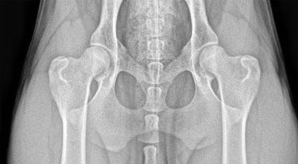
- Screenings for Hip Dysplasia are performed by a veterinarian with x-rays sent to OFA for grading and certification.
- For dogs that are between 4 months and 23 months of age, preliminary screenings are available.
- The OFA accepts preliminary consultation radiographs on puppies as young as 4 months of age for evaluation of hip conformation. If the dog is found to be dysplastic at an early age, the economic loss from the cost of training, handling, showing and so forth can be minimized and the emotional loss reduced.
- In order to publish the preliminary results:
- The dog must be at least 12 months of age at the time of radiograph
- The dog must be permanently identified via microchip or tattoo
- Note:
- Radiography of females in estrus or pregnant should be avoided due to possible increased joint laxity (subluxation) from hormonal variations. OFA recommends radiographs be taken one month after weaning pups and one month before or after a heat cycle. Physical inactivity because of illness, weather, or the owner’s management practices may also result in some degree of joint laxity. The OFA recommends evaluation when the dog is in good physical condition.
All quotations taken from:
Merck Vet Manual
OFA Website: Hip Dysplasia
OFA Website: Preliminary Evaluations
OFA Website: Hip Screening Procedures